Open Access
Peer Reviewed
ARTICLES
3205 Viewed1014 Downloaded
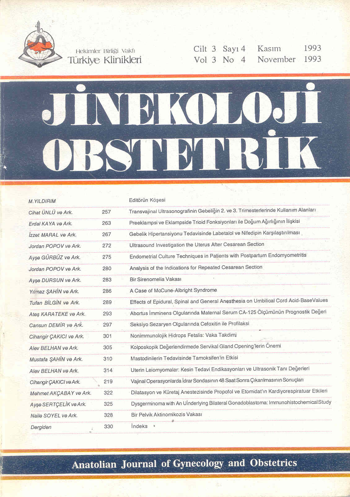
Dysgerminoma with An Underlying Bilateral Gonadoblastoma: Immunohistochemical Study
Gonadoblastoma Zemininde Gelişen Disgerminoma Olgusu: İmmunohistokimyasal Çalışma
Turkiye Klinikleri J Gynecol Obst. 1993;3(4):325-7
Article Language: TR
Copyright Ⓒ 2025 by Türkiye Klinikleri. This is an open access article under the CC BY-NC-ND license (http://creativecommons.org/licenses/by-nc-nd/4.0/)
ÖZET
Overin miks germ hücreli seks kord stromal tümörlerinden olan gonadoblastoma Scully tarafından 1953 yılında tanımlanmıştır. Bu tümörler sıklıkla disgenetik gonadlı kişilerde gözlenmekte olup, olguların çoğunda karyotipik olarak Y kromozomu mevcuttur. Biz, kliniğimizde oniki yaşında bir hastada bilateral gonadoblastoma zemininde gelişen disgerminoma saptadık. Tümöre immunohistokimyasal olarak sitokeratin ve vimentin uyguladık. Sonuçta solid adaların içindeki seks kord hücrelerinde fokal olarak vimentin ile kuvvetli, sitokeratin ile zayıf pozitif boyanma saptanmış, germ hücrelerinde ise boyanma izlenmiştir. Bu çalışmada, histopatolojik ve immunohistokimyasal bulgularımızı literatür eşliğinde tartışmayı amaçladık.
Overin miks germ hücreli seks kord stromal tümörlerinden olan gonadoblastoma Scully tarafından 1953 yılında tanımlanmıştır. Bu tümörler sıklıkla disgenetik gonadlı kişilerde gözlenmekte olup, olguların çoğunda karyotipik olarak Y kromozomu mevcuttur. Biz, kliniğimizde oniki yaşında bir hastada bilateral gonadoblastoma zemininde gelişen disgerminoma saptadık. Tümöre immunohistokimyasal olarak sitokeratin ve vimentin uyguladık. Sonuçta solid adaların içindeki seks kord hücrelerinde fokal olarak vimentin ile kuvvetli, sitokeratin ile zayıf pozitif boyanma saptanmış, germ hücrelerinde ise boyanma izlenmiştir. Bu çalışmada, histopatolojik ve immunohistokimyasal bulgularımızı literatür eşliğinde tartışmayı amaçladık.
ANAHTAR KELİMELER: Gonadoblastoma, sitokeratin, vimentin
ABSTRACT
Scully described the gonadoblastoma as a tumor of ovary composed of germ cells and sex cord derivates in 1953. The gonadoblastoma have frequently been detected in patients with gonadal dysgenesis and most of the patients whose karyotype has been determined were found to have a Y chromosome. We identified a 12 years old patient with bilateral dysgermioma developed in a gonadoblastoma. The tumor has been stained for vimentin and cytokeratin using immunohistochemical methods. It has been determined that sex cord cells in solid nests showed focal strong positive staining for vimentin and weak focal positive staining for cytokeratin and germ cells did not react to vimentin and cytokeratin. In this report, we discussed our histopathologic and immunohistochemical findings with review of the literature.
Scully described the gonadoblastoma as a tumor of ovary composed of germ cells and sex cord derivates in 1953. The gonadoblastoma have frequently been detected in patients with gonadal dysgenesis and most of the patients whose karyotype has been determined were found to have a Y chromosome. We identified a 12 years old patient with bilateral dysgermioma developed in a gonadoblastoma. The tumor has been stained for vimentin and cytokeratin using immunohistochemical methods. It has been determined that sex cord cells in solid nests showed focal strong positive staining for vimentin and weak focal positive staining for cytokeratin and germ cells did not react to vimentin and cytokeratin. In this report, we discussed our histopathologic and immunohistochemical findings with review of the literature.
MENU
POPULAR ARTICLES
MOST DOWNLOADED ARTICLES


This journal is licensed under a Creative Commons Attribution-NonCommercial-NoDerivatives 4.0 International License.


