Open Access
Peer Reviewed
ARTICLES
4128 Viewed
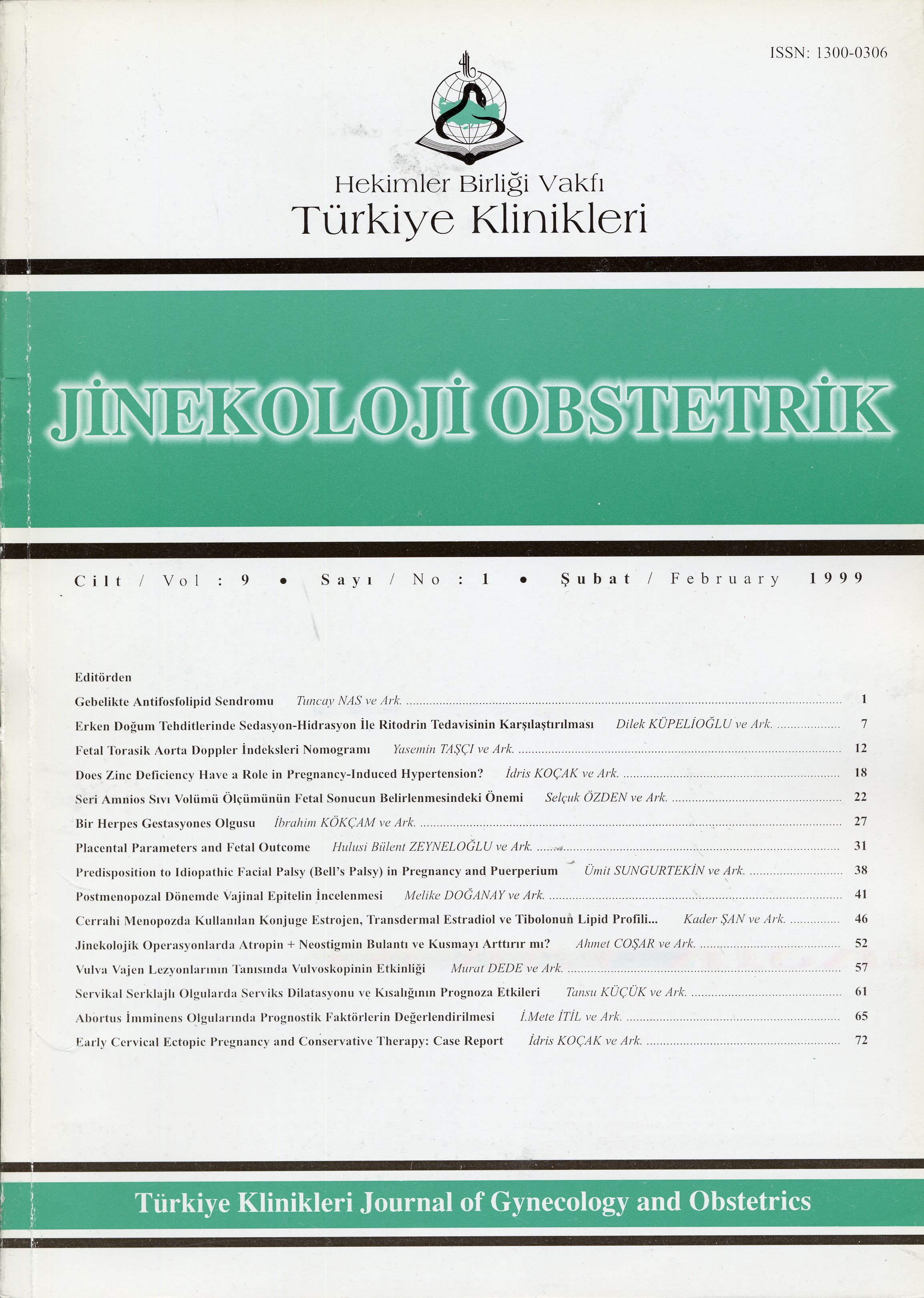
Evaluation Of The Vaginal Epithelium In The Postmenopausal Period,by Cytology, Histology And Ph Methods
Postmenopozal Dönemde Sitoloji, Histoloji ve PH Yöntemleri Kullanılarak Vajinal Epitelin İncelenmesi
Turkiye Klinikleri J Gynecol Obst. 1999;9(1):41-5
Article Language: TR
Copyright Ⓒ 2025 by Türkiye Klinikleri. This is an open access article under the CC BY-NC-ND license (http://creativecommons.org/licenses/by-nc-nd/4.0/)
ÖZET
Amaç: Postmenopozal estrojen eksikliği çalışılırken vajen epitelinin objektif olarak incelenmesi gerekmektedir. Bu çalışmada vajinal epitel değişik yöntemler ile incelenerek, atrofik ve olgun durumdaki değişiklikler araştırılmıştır. Çalışmanın Yapıldığı Yer: Dr. Zekai Tahir Burak Kadın Hastanesi, Menopoz Bölümü. Materyel ve Metod: Çalışmaya yaşları 55-62 arasında değişen doğal menopozlu 25 postmenopozal kadın alındı. Vajen epiteli estrojen öncesi ve sonrası, PH ve vajinal sitoloji yapılarak incelendi. Vajen duvarından alınan biopsiler tanımlayıcı histolojik yöntemler kullanılarak incelendi. Bulgular: Tedavi öncesi ve sonrası değerler incelendiğinde atrofiden olgun epitele geçiş gözlendi. Vajinal sitolojik maturasyon indeksi (MI) 94/6/0dan 0/65/35e doğru kayış gösterdi. Aynı şekilde vajen PHı 6.2den 4.5e doğru iniş gösterdi. Estrojen tedavisinden sonra epitel kalınlığında ve hücre tabaka sayısında artış olmuştur. Sonuç: Vajen epitelinin doğru olarak değerlendirilmesi için maturasyon indeksi ve PH kullanımı önerilmektedir.
Amaç: Postmenopozal estrojen eksikliği çalışılırken vajen epitelinin objektif olarak incelenmesi gerekmektedir. Bu çalışmada vajinal epitel değişik yöntemler ile incelenerek, atrofik ve olgun durumdaki değişiklikler araştırılmıştır. Çalışmanın Yapıldığı Yer: Dr. Zekai Tahir Burak Kadın Hastanesi, Menopoz Bölümü. Materyel ve Metod: Çalışmaya yaşları 55-62 arasında değişen doğal menopozlu 25 postmenopozal kadın alındı. Vajen epiteli estrojen öncesi ve sonrası, PH ve vajinal sitoloji yapılarak incelendi. Vajen duvarından alınan biopsiler tanımlayıcı histolojik yöntemler kullanılarak incelendi. Bulgular: Tedavi öncesi ve sonrası değerler incelendiğinde atrofiden olgun epitele geçiş gözlendi. Vajinal sitolojik maturasyon indeksi (MI) 94/6/0dan 0/65/35e doğru kayış gösterdi. Aynı şekilde vajen PHı 6.2den 4.5e doğru iniş gösterdi. Estrojen tedavisinden sonra epitel kalınlığında ve hücre tabaka sayısında artış olmuştur. Sonuç: Vajen epitelinin doğru olarak değerlendirilmesi için maturasyon indeksi ve PH kullanımı önerilmektedir.
ANAHTAR KELİMELER: Postmenopoz, Vajinal epitel, Sitoloji, Histoloji
ABSTRACT
Objective: In the study of postmenopousal vaginal oestrogen deficiency, an objective assessment of the vaginal epithelium is necessary. The present study was undertaken to evaluate different methods for assessing the vaginal epithelium, during atrophic and mature conditions. Institution: The Department of Menopouse of Dr. Zekai Tahir Burak Hospital. Materials and Methods: The research was undertaken on 25 postmenopousal women, whose ages ranged between 55-62, who were under natural menopause. The vaginal epithelium was assessed by clinical examination, PH and vaginal cytology before and after oestrogen treatment. Biopsies of the vaginal wall were studied using descriptive histology methods. Results: When comparing pre and post-treatment values from the methods, a shift towards mature values was observed, but the magnitude of the shift differed to a considerable extent. Vaginal cytology, expressed as mean maturation index (MI) shifted significantly from 94/6/0 to 0/65/35. Likewise, mean PH was significantly shifted from 6.2 to mean 4.5. The increased thickness of the epithelium and the number of cell layers could be demonstrated after oestrogen treatment. Conclusion: For objective assessment of the vaginal epithelium maturation index and PH can be recommended.
Objective: In the study of postmenopousal vaginal oestrogen deficiency, an objective assessment of the vaginal epithelium is necessary. The present study was undertaken to evaluate different methods for assessing the vaginal epithelium, during atrophic and mature conditions. Institution: The Department of Menopouse of Dr. Zekai Tahir Burak Hospital. Materials and Methods: The research was undertaken on 25 postmenopousal women, whose ages ranged between 55-62, who were under natural menopause. The vaginal epithelium was assessed by clinical examination, PH and vaginal cytology before and after oestrogen treatment. Biopsies of the vaginal wall were studied using descriptive histology methods. Results: When comparing pre and post-treatment values from the methods, a shift towards mature values was observed, but the magnitude of the shift differed to a considerable extent. Vaginal cytology, expressed as mean maturation index (MI) shifted significantly from 94/6/0 to 0/65/35. Likewise, mean PH was significantly shifted from 6.2 to mean 4.5. The increased thickness of the epithelium and the number of cell layers could be demonstrated after oestrogen treatment. Conclusion: For objective assessment of the vaginal epithelium maturation index and PH can be recommended.
MENU
POPULAR ARTICLES
MOST DOWNLOADED ARTICLES


This journal is licensed under a Creative Commons Attribution-NonCommercial-NoDerivatives 4.0 International License.


