Open Access
Peer Reviewed
ARTICLES
2983 Viewed938 Downloaded
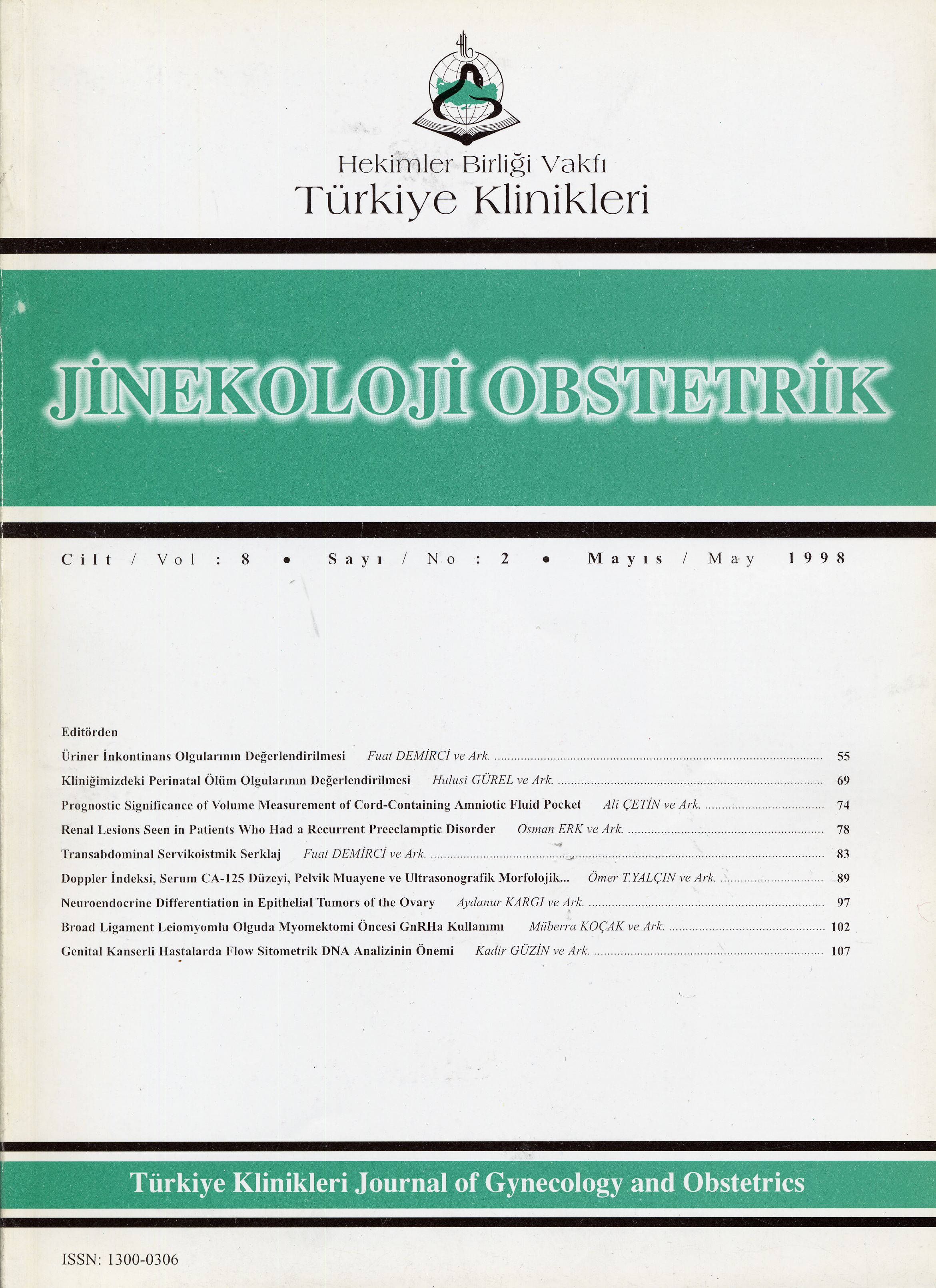
Spectrum of Renal Lesions Seen in PatientsWho Had a Recurrent Preeclamptic Disorder in Their Subsequent Pregnancies
Tekrarlayan Preeklampsi̇ Olgularinda Böbrek Lezyonlarinin Değerlendi̇ri̇lmesi̇
Turkiye Klinikleri J Gynecol Obst. 1998;8(2):78-82
Article Language: TR
Copyright Ⓒ 2025 by Türkiye Klinikleri. This is an open access article under the CC BY-NC-ND license (http://creativecommons.org/licenses/by-nc-nd/4.0/)
ÖZET
Amaç: Daha önceki gebeliklerinde de preeklampsi geçiren olguların böbreklerindeki patolojiyi saptamak ve böbrek hasarının şiddeti ile lezyonun histolojik tipi arasındaki ilişkiyi araştırmayı amaçladık. Çalışmanın yapıldığı yer: İstanbul Üniversitesi İstanbul Tıp Fakültesi Kadın Hastalıkları ve Doğum Anabilim Dalı ve İstanbul Üniversitesi İstanbul Tıp Fakültesi İç Hastalıkları Anabilim Dalı. Materyal-Metod: Daha önceki gebeliklerinde de preeklampsi geçirmiş olan ve doğumdan sonraki üç ay içinde renal patolojileri devam eden 34 preeklamptik olgu çalışma grubumuzu oluşturdu. Hastaların böbrek fonksiyonları glomerüler flitrasyon hızı ve üre klirensi ölçülerek değerlendirildi. Böbrek biyopsileri doğum sonrası üç aylık period içinde, renal patolojinin devamı durumunda yapıldı ve histopatolojik değerlendirme ışık mikroskobunda değerlendirilerek gerçekleştirildi. Bulgular: Klinik tanı olarak; 20 olguda (%58) glomerulonefrit, altı olguda (%18) kronik piyelonefrit ve sekiz olguda sadece preeklamptik toksemi saptandı. Mezangioproliferatif nefropati hem sadece preeklampsi saptanan, hemde kronik renal lezyon bulunan olgularda, en fazla görülen histopatolojik tanı oldu. Sonuç: Doğum sonrası renal patolojileri devam eden preeklamptik olgularda en sık görülen histopatolojik tanı "Mezangioproliferatif Nefropati" olarak belirlendi. Tanıda klinik, laboratuar ve gerekirse histopatolojik tanının önemi vurgulandı.
Amaç: Daha önceki gebeliklerinde de preeklampsi geçiren olguların böbreklerindeki patolojiyi saptamak ve böbrek hasarının şiddeti ile lezyonun histolojik tipi arasındaki ilişkiyi araştırmayı amaçladık. Çalışmanın yapıldığı yer: İstanbul Üniversitesi İstanbul Tıp Fakültesi Kadın Hastalıkları ve Doğum Anabilim Dalı ve İstanbul Üniversitesi İstanbul Tıp Fakültesi İç Hastalıkları Anabilim Dalı. Materyal-Metod: Daha önceki gebeliklerinde de preeklampsi geçirmiş olan ve doğumdan sonraki üç ay içinde renal patolojileri devam eden 34 preeklamptik olgu çalışma grubumuzu oluşturdu. Hastaların böbrek fonksiyonları glomerüler flitrasyon hızı ve üre klirensi ölçülerek değerlendirildi. Böbrek biyopsileri doğum sonrası üç aylık period içinde, renal patolojinin devamı durumunda yapıldı ve histopatolojik değerlendirme ışık mikroskobunda değerlendirilerek gerçekleştirildi. Bulgular: Klinik tanı olarak; 20 olguda (%58) glomerulonefrit, altı olguda (%18) kronik piyelonefrit ve sekiz olguda sadece preeklamptik toksemi saptandı. Mezangioproliferatif nefropati hem sadece preeklampsi saptanan, hemde kronik renal lezyon bulunan olgularda, en fazla görülen histopatolojik tanı oldu. Sonuç: Doğum sonrası renal patolojileri devam eden preeklamptik olgularda en sık görülen histopatolojik tanı "Mezangioproliferatif Nefropati" olarak belirlendi. Tanıda klinik, laboratuar ve gerekirse histopatolojik tanının önemi vurgulandı.
ANAHTAR KELİMELER: Toksemi, Kronik renal hastalık, Mezangioproliferatif nefropati
ABSTRACT
Objective: To evaluate the renal lesions seen in the patients who had a recurrent preeclamptic disorder in their subsequent pregnancies and to asses the correlation between the histological type of lesions and the severity of the renal disease. Institution: Istanbul University Istanbul Medical Faculty, Department of Internal Medicine and Department of Obstetrics and Gynecology. Material and metods: 34 women with the signs of severe preeclampsia during their previous pregnancies, have been studied. Kidney functions were determined by GFR and urea clearance. Renal biopsies were done within three month, after delivery if there are the hypertensive disorders or/and proteinuria after this time. All tissues were processed for examination under light microscopy. Results: Among 34 patients with the clinical sign and symptoms of toxemia a biopsy diagnosis of underlying glomerulonephritis had been made in 20 cases ( 58 % ), chronic pyelonephritis in 6 cases ( 18 % ) and pure preeclamptic toxemia in 8 cases (24%). Mesangioproliferative nephropathia have been found to be the most common histological lesions seen in both pure toxemia (87%) and chronic renal lesions (60%). Conclusion: Mesangioproliferative nephropathia was found to be the most common renal lesions seen in both pure toxemia and chronic renal disease. Significance of constellation of clinical, laboratory and histological findings in making the find diagnosis have been emphasized.
Objective: To evaluate the renal lesions seen in the patients who had a recurrent preeclamptic disorder in their subsequent pregnancies and to asses the correlation between the histological type of lesions and the severity of the renal disease. Institution: Istanbul University Istanbul Medical Faculty, Department of Internal Medicine and Department of Obstetrics and Gynecology. Material and metods: 34 women with the signs of severe preeclampsia during their previous pregnancies, have been studied. Kidney functions were determined by GFR and urea clearance. Renal biopsies were done within three month, after delivery if there are the hypertensive disorders or/and proteinuria after this time. All tissues were processed for examination under light microscopy. Results: Among 34 patients with the clinical sign and symptoms of toxemia a biopsy diagnosis of underlying glomerulonephritis had been made in 20 cases ( 58 % ), chronic pyelonephritis in 6 cases ( 18 % ) and pure preeclamptic toxemia in 8 cases (24%). Mesangioproliferative nephropathia have been found to be the most common histological lesions seen in both pure toxemia (87%) and chronic renal lesions (60%). Conclusion: Mesangioproliferative nephropathia was found to be the most common renal lesions seen in both pure toxemia and chronic renal disease. Significance of constellation of clinical, laboratory and histological findings in making the find diagnosis have been emphasized.
MENU
POPULAR ARTICLES
MOST DOWNLOADED ARTICLES


This journal is licensed under a Creative Commons Attribution-NonCommercial-NoDerivatives 4.0 International License.


