Open Access
Peer Reviewed
ARTICLES
3444 Viewed1220 Downloaded
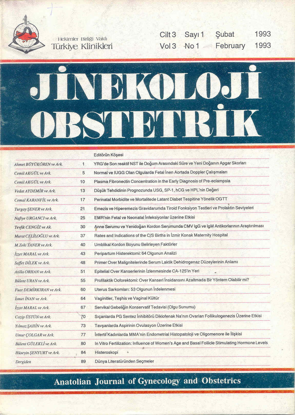
Vaginitis, Diagnosis And Vaginal Culture
Vaginitler, Teşhis ve Vaginal Kültür
Turkiye Klinikleri J Gynecol Obst. 1993;3(1):64-6
Article Language: TR
Copyright Ⓒ 2025 by Türkiye Klinikleri. This is an open access article under the CC BY-NC-ND license (http://creativecommons.org/licenses/by-nc-nd/4.0/)
ÖZET
Vaginal akıntıların teşhis ve tedavisini daha etkin kılmak için vaginal kültür alınması uygundur. Vaginal kültürü daha önce kuru steril tüple alıp laboratuvara yollarken, şimdi 1.si içinde 0.5 cc. Serum fizyolojik olan tüple, 2.si içinde stuart besiyeri olan tüple taşıyarak çeşitli besiyerlerine ekildi. Çalışma polikliniğe vaginal akıntı şikayeti ile başvuran 140 olgu üzerinde yapıldı. Olgular 2. gruba ayrıldı. I. Grup: 84 olguda materyel kuru pamuklu ekiviyon çubuğu ile alındı ve kuru steril tüple laboratuvara ulaştırıldı. II. Grup: 56 olguda ise alınan materyel birincisi-stuart transport besiyerine, ikincisi 0.5 cc. Serum fizyolojik içeren tüpe alındı ve uygun vasatlara ekim yapıldı. II. Grupta normal flora sayısı azaldı. Gardnerella ve Candida sayısında artma meydana geldi. Taşıma vasatları kullanmak ve uygun vasatlara ekim yapmak kültür neticesini müspet olarak etkiledi. CNA vasatı ve stuart taşıma vasatı, kullanılınca G. Vaginalisin kültürde bulunma nispeti %7.1'den %21.6'ya artış gösterdi (x2=5.5008>x2, t=3.841). Ve istatistiksel fark önemli idi.
Vaginal akıntıların teşhis ve tedavisini daha etkin kılmak için vaginal kültür alınması uygundur. Vaginal kültürü daha önce kuru steril tüple alıp laboratuvara yollarken, şimdi 1.si içinde 0.5 cc. Serum fizyolojik olan tüple, 2.si içinde stuart besiyeri olan tüple taşıyarak çeşitli besiyerlerine ekildi. Çalışma polikliniğe vaginal akıntı şikayeti ile başvuran 140 olgu üzerinde yapıldı. Olgular 2. gruba ayrıldı. I. Grup: 84 olguda materyel kuru pamuklu ekiviyon çubuğu ile alındı ve kuru steril tüple laboratuvara ulaştırıldı. II. Grup: 56 olguda ise alınan materyel birincisi-stuart transport besiyerine, ikincisi 0.5 cc. Serum fizyolojik içeren tüpe alındı ve uygun vasatlara ekim yapıldı. II. Grupta normal flora sayısı azaldı. Gardnerella ve Candida sayısında artma meydana geldi. Taşıma vasatları kullanmak ve uygun vasatlara ekim yapmak kültür neticesini müspet olarak etkiledi. CNA vasatı ve stuart taşıma vasatı, kullanılınca G. Vaginalisin kültürde bulunma nispeti %7.1'den %21.6'ya artış gösterdi (x2=5.5008>x2, t=3.841). Ve istatistiksel fark önemli idi.
ANAHTAR KELİMELER: Vaginitis, vaginal kültür, candida, trikomonas vaginalis
ABSTRACT
Vaginal samples should be taken and cultured to diagnose and to cure the vaginal infections more effectively. The samples of the vaginal discharge was taken by immersing a swab and it is put in dry tube for the laboratory tests previously. Now, we use two tubes, the first tube contains the saline solution of 0.5 cc and the second tube contains the stuart media and we transport these tubes to laboratory for plating the selective media. We studied on 140 cases that have vaginal discharge. The cases are divided into two groups. On the first group which contains 84 cases, the sample of the vaginal discharge with immersing a swab and transport to the laboratory in a dry sterile tube. On the second group which contains 56 cases, the vaginal discharge samples are put in two different tubes. One of the tubes contains the stuart media and the other one contains 0.5 cc saline solution and the specimens in the tubes are plated on the appropriate media. The amount of Gardnerella vaginalis and vaginal candidiasis increased whereas the amount of normal flora decreased with the new method. Using of the new carrying media and plating on the appropriate media affected the result of the culture specimen positively. When CNA media (Difco) is used for G vaginalis and stuart media to transport, the percentage of G. Vaginalis increased from 7.1% to 21.6% that is formulated as (x2=5.5008>x2, t=3.841) an important amount.
Vaginal samples should be taken and cultured to diagnose and to cure the vaginal infections more effectively. The samples of the vaginal discharge was taken by immersing a swab and it is put in dry tube for the laboratory tests previously. Now, we use two tubes, the first tube contains the saline solution of 0.5 cc and the second tube contains the stuart media and we transport these tubes to laboratory for plating the selective media. We studied on 140 cases that have vaginal discharge. The cases are divided into two groups. On the first group which contains 84 cases, the sample of the vaginal discharge with immersing a swab and transport to the laboratory in a dry sterile tube. On the second group which contains 56 cases, the vaginal discharge samples are put in two different tubes. One of the tubes contains the stuart media and the other one contains 0.5 cc saline solution and the specimens in the tubes are plated on the appropriate media. The amount of Gardnerella vaginalis and vaginal candidiasis increased whereas the amount of normal flora decreased with the new method. Using of the new carrying media and plating on the appropriate media affected the result of the culture specimen positively. When CNA media (Difco) is used for G vaginalis and stuart media to transport, the percentage of G. Vaginalis increased from 7.1% to 21.6% that is formulated as (x2=5.5008>x2, t=3.841) an important amount.
MENU
POPULAR ARTICLES
MOST DOWNLOADED ARTICLES


This journal is licensed under a Creative Commons Attribution-NonCommercial-NoDerivatives 4.0 International License.


