Open Access
Peer Reviewed
ORIGINAL RESEARCH
3136 Viewed1157 Downloaded
Contralateral Uterine Tube Ultrastructures In Adnexial Torsion
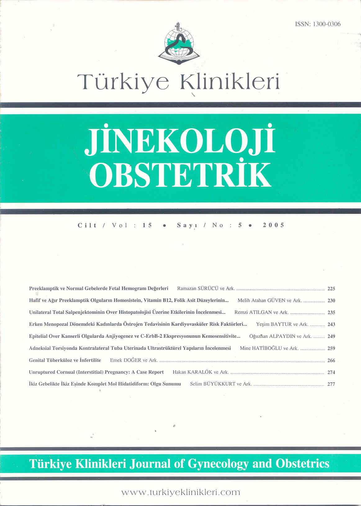
Adneksial Torsiyonda Kontralateral Tuba Uterinada Ultrastrüktürel Yapıların İncelenmesi
Turkiye Klinikleri J Gynecol Obst. 2005;15(5):259-65
Article Language: TR
Copyright Ⓒ 2025 by Türkiye Klinikleri. This is an open access article under the CC BY-NC-ND license (http://creativecommons.org/licenses/by-nc-nd/4.0/)
ÖZET
Amaç: Unilateral adneksa torsiyonunda kontralateral tuba uterinanın etkilenip etkilenmediğini araştırmak amacıyla tavşanlarda deneysel çalışma yapıldı. Gereç ve Yöntemler: Çalışmada 19 adet Yeni Zelanda cinsi, albino, dişi tavşan kullanıldı. Tüm deneklerde menstrual siklusun tuba uterinaya olan etkileri serum gonodotropin (PMSG) ile senkronize edildi. Kontrol grubuna sadece laparatomi yapılıp (sham ameliyat), ikinci grubun 12 saat, üçüncü grubun 24 saat süreyle sol adneksaları torsiyone edildi ve süre sonunda kontralateral tuba uterinalar eksize edildi. Bulgular: Kontrol, 12. ve 24. saat iskemi gruplarının ışık mikroskopik ve elektron mikroskopik bulguları karşılaştırıldı. Salgı hücreleri, salgı granülleri ve salgı aktiviteleri 12 saat iskemide artmış olmasına karşın 24 saatlik iskemide azaldığı gözlendi. Kinosilyumlu epitel hücrelerinde ise iskemi süresi ile orantılı artış gözlendi. 24 saatte kontralateral tuba uterinada lenfositik infiltrasyon görüldü. Sonuç: Epitel yapısındaki değişimin muhtemelen kan akımı ve hormonal değişime bağlı geliştiği, lenfositik infiltrasyon ise hücresel immünitenin aktivasyonunu düşündürmüştür.
Amaç: Unilateral adneksa torsiyonunda kontralateral tuba uterinanın etkilenip etkilenmediğini araştırmak amacıyla tavşanlarda deneysel çalışma yapıldı. Gereç ve Yöntemler: Çalışmada 19 adet Yeni Zelanda cinsi, albino, dişi tavşan kullanıldı. Tüm deneklerde menstrual siklusun tuba uterinaya olan etkileri serum gonodotropin (PMSG) ile senkronize edildi. Kontrol grubuna sadece laparatomi yapılıp (sham ameliyat), ikinci grubun 12 saat, üçüncü grubun 24 saat süreyle sol adneksaları torsiyone edildi ve süre sonunda kontralateral tuba uterinalar eksize edildi. Bulgular: Kontrol, 12. ve 24. saat iskemi gruplarının ışık mikroskopik ve elektron mikroskopik bulguları karşılaştırıldı. Salgı hücreleri, salgı granülleri ve salgı aktiviteleri 12 saat iskemide artmış olmasına karşın 24 saatlik iskemide azaldığı gözlendi. Kinosilyumlu epitel hücrelerinde ise iskemi süresi ile orantılı artış gözlendi. 24 saatte kontralateral tuba uterinada lenfositik infiltrasyon görüldü. Sonuç: Epitel yapısındaki değişimin muhtemelen kan akımı ve hormonal değişime bağlı geliştiği, lenfositik infiltrasyon ise hücresel immünitenin aktivasyonunu düşündürmüştür.
ANAHTAR KELİMELER: Adneksa, torsiyon, tuba uterina, ultrastrüktür, elektron mikroskopisi
ABSTRACT
Objective: To evaluate the tissue structure of contralateral uterine tuba in case of unilateral adnexial torsion an experimental study was setup. Material and Methods: Nineteen adult, albino, New Zelland rabbits were used. The effects of menstrual cycle on tuba uterina were synchronised by serum gonadotropin (PMSG). Animals in the control group were subjected to laparatomy only (Sham operation). Adnexia of the animals were subjected to torsion for a period of 12 hours in the second group and for 24 hours in the third group. Following these interventions contralateral uterine tubes were excised in all animals. Results: Histological findings on light and electron microscopy of the groups in comparison with each other revealed decrease in secretory cells, their granules and activity at 24 hours ischemia; though initial rise were detected at 12 hours ischemia. Ciliated cells of contralateral uterine tube epithelium increased in number with respect to the duration of the unilateral adnexial ischemia. At 24 hours ischemia group lymphocytes were observed as infiltrated to the contralateral uterine tubes. Conclusion: Epitelial changes were possibly due to blood flow and hormonal alterations and lenfositic infiltration was a possible remark of the activation of the cellular immunity.
Objective: To evaluate the tissue structure of contralateral uterine tuba in case of unilateral adnexial torsion an experimental study was setup. Material and Methods: Nineteen adult, albino, New Zelland rabbits were used. The effects of menstrual cycle on tuba uterina were synchronised by serum gonadotropin (PMSG). Animals in the control group were subjected to laparatomy only (Sham operation). Adnexia of the animals were subjected to torsion for a period of 12 hours in the second group and for 24 hours in the third group. Following these interventions contralateral uterine tubes were excised in all animals. Results: Histological findings on light and electron microscopy of the groups in comparison with each other revealed decrease in secretory cells, their granules and activity at 24 hours ischemia; though initial rise were detected at 12 hours ischemia. Ciliated cells of contralateral uterine tube epithelium increased in number with respect to the duration of the unilateral adnexial ischemia. At 24 hours ischemia group lymphocytes were observed as infiltrated to the contralateral uterine tubes. Conclusion: Epitelial changes were possibly due to blood flow and hormonal alterations and lenfositic infiltration was a possible remark of the activation of the cellular immunity.
MENU
POPULAR ARTICLES
MOST DOWNLOADED ARTICLES


This journal is licensed under a Creative Commons Attribution-NonCommercial-NoDerivatives 4.0 International License.


