Open Access
Peer Reviewed
ARTICLES
3055 Viewed979 Downloaded
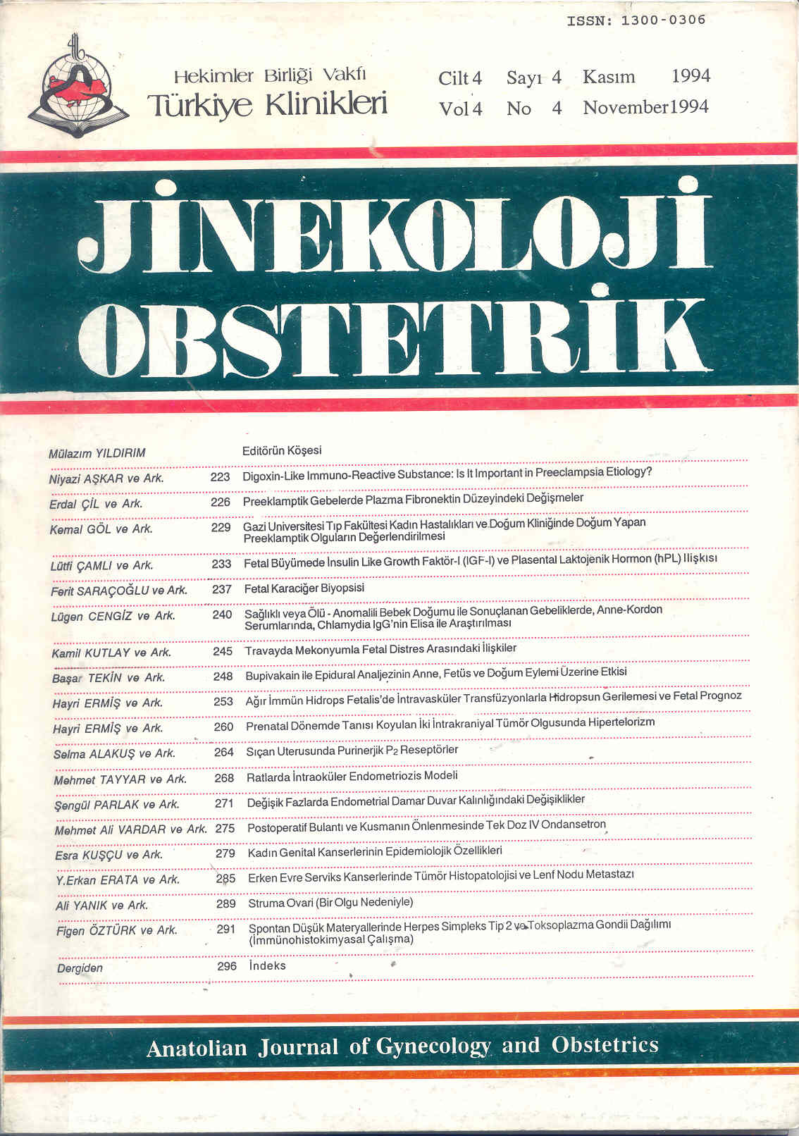
Intraocular Endometriosis In Rats
Ratlarda İntraoküler Endometriozis Modeli
Turkiye Klinikleri J Gynecol Obst. 1994;4(4):268-70
Article Language: TR
Copyright Ⓒ 2025 by Türkiye Klinikleri. This is an open access article under the CC BY-NC-ND license (http://creativecommons.org/licenses/by-nc-nd/4.0/)
ÖZET
Amaç: Endometriozis etyoloji ve tedavisini incelerken direkt gözlem yapılabilecek bir hayvan modeli oluşturmak. Çalışmanın Yapıldığı Yer: Erciyes Üniversitesi Tıp Fakültesi Kadın Hastalıkları ve Doğum AD, Kayseri. Materyel ve Metod: Ortalama ağırlıkları 200 g olan 8 dişi Swiss-Albino rat kullanıldı. Eter anestezisini takiben steril şartlarda laparotomi yapılarak uterin horndan bir parça eksize edildi. Alınan implant aynı seansta gözün iris tabakası üzerine yerleştirildi. Altı hafta süreyle göz bulguları incelendi. Bu sürenin sonunda gözler enüklee edilerek histopatolojik inceleme yapıldı. Bulgular: Tüm gözlerde, ön kamerada uterin implantları varlığı ve büyüdüğü gözlendi. Histopatolojik incelemede tüm gözlerde iyi damarlanmış endometrial yapılar ve hemosiderin birikimi izlendi. Sonuç: İntraoküler endometriozis modelinin direkt gözlem ve takip yapabilme avantajı nedeniyle endometriozis modeli olarak kullanılabileceği kanısına vardık.
Amaç: Endometriozis etyoloji ve tedavisini incelerken direkt gözlem yapılabilecek bir hayvan modeli oluşturmak. Çalışmanın Yapıldığı Yer: Erciyes Üniversitesi Tıp Fakültesi Kadın Hastalıkları ve Doğum AD, Kayseri. Materyel ve Metod: Ortalama ağırlıkları 200 g olan 8 dişi Swiss-Albino rat kullanıldı. Eter anestezisini takiben steril şartlarda laparotomi yapılarak uterin horndan bir parça eksize edildi. Alınan implant aynı seansta gözün iris tabakası üzerine yerleştirildi. Altı hafta süreyle göz bulguları incelendi. Bu sürenin sonunda gözler enüklee edilerek histopatolojik inceleme yapıldı. Bulgular: Tüm gözlerde, ön kamerada uterin implantları varlığı ve büyüdüğü gözlendi. Histopatolojik incelemede tüm gözlerde iyi damarlanmış endometrial yapılar ve hemosiderin birikimi izlendi. Sonuç: İntraoküler endometriozis modelinin direkt gözlem ve takip yapabilme avantajı nedeniyle endometriozis modeli olarak kullanılabileceği kanısına vardık.
ANAHTAR KELİMELER: İntraoküler endometriozis, model, rat
ABSTRACT
Objective: To find an animal model which can be used to investigate the etiology and the treatment of endometriosis on which direct observation is possible. Institution: Erciyes University Faculty of Medicine, Department of Obstetrics and Gynecology. Material and Methods: Eight female Swiss-Albino rats weighing 200 g on average were used. After ether anesthesia, laparotomy was performed and a piece of uterine horn was excised under sterile conditions. The excised implants were placed on the iris of the eyes in the same surgical proces. Findings of the eyes have been investigated for 6 weeks. At the end of this period, eyes were enucleated and histopathological procedures were performed. Findings: It was observed that all the implants were existing and growing in all anterior cameras of the eyes. In histopathological investigation, well vasculized endometrial developments and the collection of hemosiderin were seen in all eyes. Conclusion: We think that intraocular endometriosis model can be used as an endometriosis model because of its advantages as direct observation and follow-up.
Objective: To find an animal model which can be used to investigate the etiology and the treatment of endometriosis on which direct observation is possible. Institution: Erciyes University Faculty of Medicine, Department of Obstetrics and Gynecology. Material and Methods: Eight female Swiss-Albino rats weighing 200 g on average were used. After ether anesthesia, laparotomy was performed and a piece of uterine horn was excised under sterile conditions. The excised implants were placed on the iris of the eyes in the same surgical proces. Findings of the eyes have been investigated for 6 weeks. At the end of this period, eyes were enucleated and histopathological procedures were performed. Findings: It was observed that all the implants were existing and growing in all anterior cameras of the eyes. In histopathological investigation, well vasculized endometrial developments and the collection of hemosiderin were seen in all eyes. Conclusion: We think that intraocular endometriosis model can be used as an endometriosis model because of its advantages as direct observation and follow-up.
MENU
POPULAR ARTICLES
MOST DOWNLOADED ARTICLES


This journal is licensed under a Creative Commons Attribution-NonCommercial-NoDerivatives 4.0 International License.


